Team:Oxford/Our solution

Our overall design incorporates both sensitive C. difficile detection and targeted killing.

C. difficile uses quorum signalling to coordinate high population density with toxin synthesis. Such toxins, notably TcdA and TcdB1, cause inflammation of the gut lining and consequently the symptoms of C. difficile infection (CDI). Such quorum signaling molecules thus provide an ideal biomarker for our detection system. Detection of these molecules (as opposed to the toxins) should allow our ProQuorum system to detect high-density colonies of C. difficile before significant toxin release.

Detection/Regulatory System

C. difficile produces the quorum signalling molecule, specifically referred to as the auto-inducer peptide (AIP) via two membrane proteins AgrB and AgrD, with the latter being modified to form AIP. This AIP is then secreted into the extracellular media.

Thus, for our ProQuorum system, we make use of C. difficile’s own detection system for AIP. This is a two-component detection system consisting of the two proteins AgrA and AgrC. AgrC acts as a specific transmembrane receptor for AIP and upon ligand binding, the activated receptor causes phosphorylation and subsequent dimerisation of AgrA. This dimer then acts as a specific transcription factor to activate the Agr promoter.2

Detection/Regulatory System

Endolysin/Expression System

The activation of the Agr promoter will induce the expression of the C. difficile-specific CD27L endolysin, derived from the bacteriophage ΦCD27L.3 The endolysin is subsequently secreted via the Sec pathway courtesy of its slpMOD secretion tag. This tag has been specifically developed for use in the commensal gut bacterium Lactococcus lactis4 and Lactobacillus johnsonii.5 Thus, it provides a suitable secretion tag for our chassis of Lactobacillus reuteri.

The CD27L endolysin, as an N-acetylmuramoyl-L-alanine amidase6, specifically recognises glycopeptides of the C. difficile cell wall and cleaves amide bonds to cause degradation of the peptidoglycan cell wall. The resulting cell undergoes osmotic lysis. Such a choice of drug molecule enables rapid and targeted lysis of pathogenic C. difficile cells whilst preserving the remainder of the gut microbiome.

Expression/Secretion System

This overall system should enable both the specific detection and targeted killing of C. difficile colonies, providing substantial improvements over the broad-spectrum antibiotics metronidazole and vancomycin1 currently used to treat CDI.

Overall System

Our Design Process

We ultimately achieved this design not just by ourselves, but with huge contributions from various parties including our mathematical modelling team, academic experts and previous CDI patients. Further detail can be found in our Modelling, Human Practices and Public Engagement pages.

In order to achieve maximal efficacy of treatment, we wanted our system to function directly at the site of infection: the gut. This had a significant influence on our choice of chassis. Resulting from discussions with Dr David Eyre, a C. difficile expert, who first suggested that we explore Lactobacillus strains, as these bacteria are already well adapted to grow in the human gut. We then decided to use L. reuteri as our chassis due to its intrinsic resistance to metrodinazole, vanocymin and fidoamicin 7 and its ability to secrete reuterin, a broad spectrum antibiotic when in the presence of glycerol. Together, these tools make wild-type L. reuteri an effective C. difficile suppressant. Our literature review5,7,9 supported these claims and we decided to use the probiotic strain L. reuteri, which has been shown to act as an anti-microbial probiotic7 and fulfills all our design criteria. It is also commonly used as a safe-to-consume probiotic as mentioned by Mu et al. (2018).9

Additional advantages of L. reuteri as our chassis include its fulfilment of our design criteria, including its resistance to our CD27L endolysin and its Gram-positive nature (like C. difficile) which should enable functional expression of the transmembrane detection receptor AgrC.

Our final design choice was the particular plasmid expression vector for L. reuteri. As per Lizier et al. (2010)8, we chose pTRKH3 as it had be used effectively in L. reuteri work.

>

References

# Reference
1 Smits, Wiep Klaas, et al. “Clostridium Difficile Infection.” Nature Reviews Disease Primers, vol. 2, no. 1, July 2016, doi:10.1038/nrdp.2016.20.
2 Darkoh, Charles, et al. “Toxin Synthesis by Clostridium Difficile Is Regulated through Quorum Signaling.” MBio, Feb. 2015.
3 Mayer, M. J., et al. “Molecular Characterization of a Clostridium Difficile Bacteriophage and Its Cloned Biologically Active Endolysin.” Journal of Bacteriology, vol. 190, no. 20, 2008, pp. 6734–6740., doi:10.1128/jb.00686-08.
4 Fernandez, A., et al. “Enhanced Secretion of Biologically Active Murine Interleukin-12 by Lactococcus Lactis.” Applied and Environmental Microbiology, vol. 75, no. 3, May 2008, pp. 869–871., doi:10.1128/aem.01728-08.
5 Gervasi, Teresa, et al. “Expression and Delivery of an Endolysin to Combat Clostridium Perfringens.” Applied Microbiology and Biotechnology, vol. 98, no. 6, 2013, pp. 2495–2505., doi:10.1007/s00253-013-5128-y.
6 Mayer, M. J., et al. “Structure-Based Modification of a Clostridium Difficile-Targeting Endolysin Affects Activity and Host Range.” Journal of Bacteriology, vol. 193, no. 19, 2011, pp. 5477–5486., doi:10.1128/jb.00439-11.
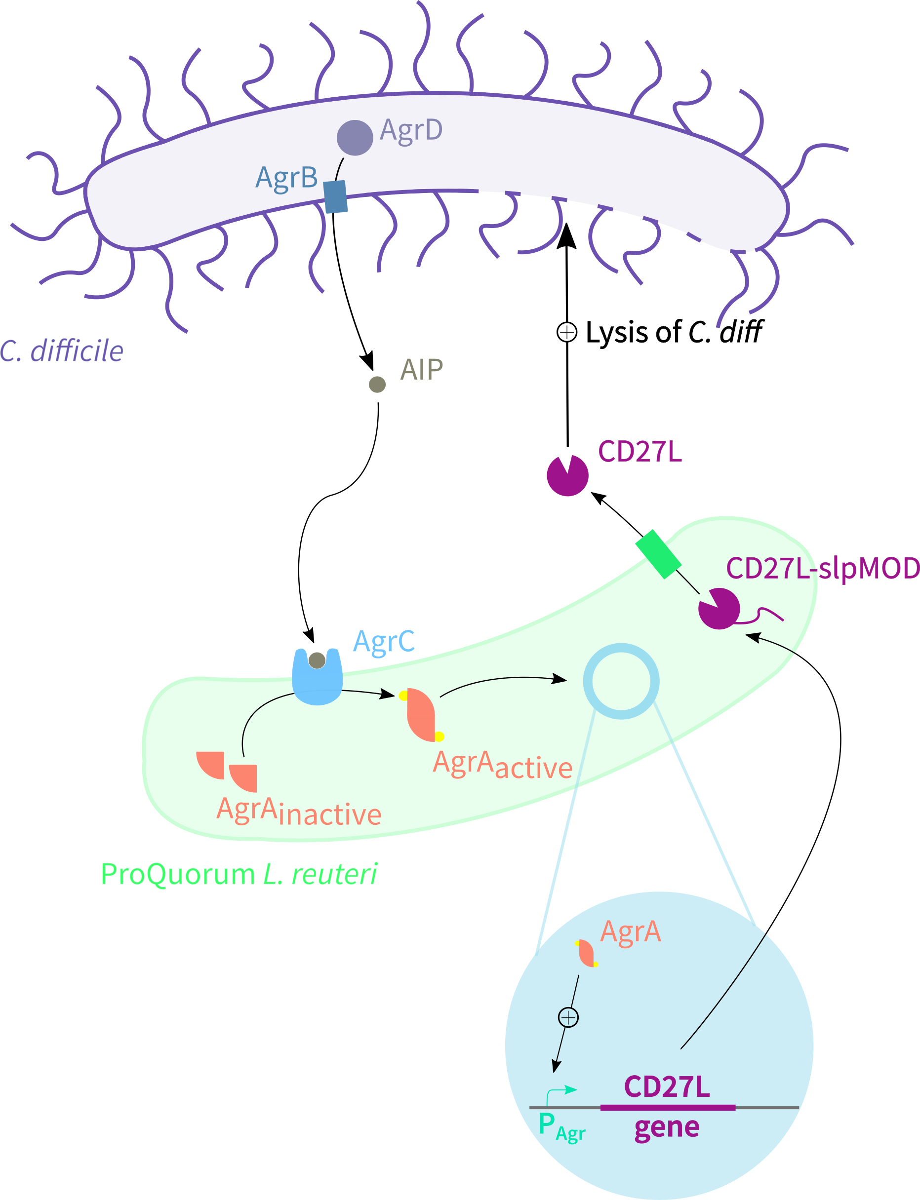
7 Spinler, Jennifer K., et al. “Next-Generation Probiotics Targeting Clostridium Difficile through Precursor-Directed Antimicrobial Biosynthesis.” Infection and Immunity, vol. 85, no. 10, 2017, doi:10.1128/iai.00303-17.
8 Lizier, Michela, et al. “Comparison of Expression Vectors in Lactobacillus Reuteri Strains.” FEMS Microbiology Letters, vol. 308, no. 1, Aug. 2010, pp. 8–15., doi:10.1111/j.1574-6968.2010.01978.x.
9 Mu, Qinghui, et al. “Role of Lactobacillus Reuteri in Human Health and Diseases.” Frontiers in Microbiology, vol. 9, 2018, doi:10.3389/fmicb.2018.00757.